Medical Microbiology

Thursday, December 14, 2006

3rd Entry - Summary of all Cases

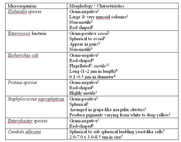
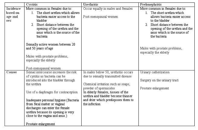
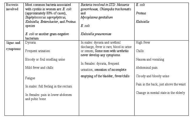
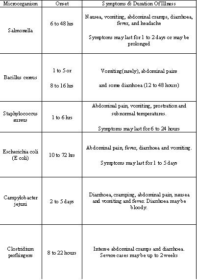
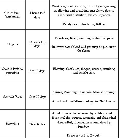
All the necessary information is collated by all members and organised into tables. This can be seen below.
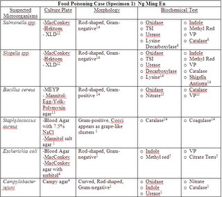


Abbreviations:
IMVic: indole-methyl red-Voges-Proskeur-Citrate
ONPG: O-nitrophenyl-β-D-galactopyranoside
OF: oxidation-fermentation
TSI: triple sugar iron
VP: Voges-Proskeur
MR: Methyl Red

References:
(1) http://findarticles.com/>
(2) http://medinfo.ufl.edu>
(3) http://tucom.bigbri.net/>
(4) http://en.wikipedia.org/>
(5) http://www.umm.edu/>
(6) http://textbookofbacteriology.net>
(7) Gabriel Virella. Microbiology and infectious diseases. Third edition
(8) http://w3.ouhsc.edu/>
(9)http://www.microbelibrary.org>
(10) http://members.tripod.com
(11) http://www.mc.maricopa.edu/>
(12) http://www.who.int/>
(13) http://www.rci.rutgers.edu/>
(14) G.Brooks, J.Butel and S.Morse. (2004). Jawetz, Melnick & Adelberg’s Medical Microbiology
(15) Henry D. Isenberg. Essential procedures for clinical microbiology

Friday, December 08, 2006

Culture media required for microorganism growth

As we will have to prepare the media culture for growing of the possible microorganisms, we have gathered information on the possible microorganisms and its corresponding appropriate culture media. This is summarised in the tables below.

For UTI cases (Patient 1,3 and 5) – Urine specimen [information gathered by Syafiqah & Samantha]


For enterocolitis case (patient 2 -Kwan Siew Lan ) [information gathered by Yasmin]


For food poisoning case (patient 4 - Ng Ming En) [8]
[information gathered by Farhana]


References:
[1] G.Brooks, J.Butel and S.Morse. (2004). Jawetz, Melnick & Adelberg’s Medical Microbiology
[2] http://medinfo.ufl.edu/year2/mmid/bms5300/bugs/klebpne.html
[3] http://en.wikipedia.org/wiki/Enterobacter
[4] http://medinfo.ufl.edu/year2/mmid/bms5300/bugs/candalb.html
[5] http://medinfo.ufl.edu/year2/mmid/bms5300/bugs/esccoli.html
[6] http://medic.med.uth.tmc.edu/path/bile.htm
[7] http://www.microbelibrary.org/ASMOnly/details.asp?id=2315&Lang
[8] http://www.findarticles.com/p/articles/mi_gGENH/is_/ai_2699003751

Thursday, December 07, 2006

Patient 1: Khong fay fay (2nd entry)

Reasons why cystitis was chosen for the diagnosis

1. Nausea and vomiting are signs and symptoms of pyelonephritis
a. Nausea and vomiting were not observed in this patient [1]

2. Causes of pyelonephritis include [1]
a. Urinary catherization[Kidney Infection (Pyelonephritis)
b. Surgery on urinary tract
c. Physical obstruction to the flow of urine due to a prostate enlargement or kidney stone or the backflow of urine from the bladder into the ureters increases the risk of pyelonephritis

However,
à Urinary catherization was not performed in this patient
à No history of surgery was indicated
à Patient is a female, hence physical obstruction to the flow of urine due to prostate enlargment is unlikely

3. Pyelonephritis is usually a spread of bacterial infection from the bladder [2]
à Risk of infection increases if there is a history of cystitis
à Hence even if pyelonephritis was present, cystitis is still the primary diagnosis

4. Pyelonephritis occurs less commonly in comparison to cystits. [3]

Complicated and Uncomplicated Cystitis
Cystitis is a common female problem. About 90% of UTIs in women are uncomplicated but recurrent. [4]

Cystitis is referred to as complicated if there are urologic abnormalities such as urinary retention, urinary stones and post surgery [5]

Bacteria found in cystitis
Uncomplicated cystitis
1. E. coli is the most commonly isolated microorganism in 80 - 90% of cystitis in young women followed by
2. Staphylococcus saprophyticus
3. Enterococci

Bacteria such as
1. Klebsiella
2. Proteus
3. Pseudomonads
constitute a lower percentage of infecting agents in uncomplicated cystits

Complicated cystitis
The spectrum of infecting bacteria in complicated cystitis is broader. Common infecting agents include
1. E. coli
2. Klebsiella
3. Proteus
4. Enterobacter
5. Enterococci
6. Pseudomonads
7. Staphylococci

Conclusion
Since uncomplicated cystitis is more common, the most probable bacteria that can be isolated from the bladder of this patient includes E. coli, Staphylococcus saprophyticus and Enterococci. Now that the common microorganisms have been identified, the next step is to find out ways to isolate the following bacteria and the biochemical tests that can be used for the identification of these bacteria.

References
1. http://www.merck.com/mmhe/sec11/ch149/ch149e.html
2. http://www.intelihealth.com/IH/ihtIH/WSIHW000/9339/10227.html
3. http://www.nlm.nih.gov/medlineplus/ency/article/000522.htm
4. http://www.faqs.org/health/Sick-V1/Cystitis.html
5. http://www.touchbriefings.com/pdf/1134/riedl.pdf
6. G.Brooks, J.Butel and S.Morse. (2004). Jawetz, Melnick & Adelberg’s Medical Microbiology

Patient 3: Maisy Wong (2nd entry)

Complaints (key words): Fever, chills, bladder distension. Patient is on indwelling catheter.

In this case, the urinary tract infection (UTI) can be caused by several reasons:

1) The patient is a 66 years old female. Hence the UTI may also be due to post menopause.
It is said that ‘the risk for UTI, both symptomatic and asymptomatic, is highest in women after menopause’ [2]. This may be due to bladder or uterine prolapse leading to incomplete bladder emptying [4]. Menopause may cause biologic changes. This in turn ‘put older women at particular risk for primary and recurring UTIs’ [2].

Post menopausal women will encounter estrogen loss. With this, the walls of urinary tract will thin out hence weakening the mucous membrane and reducing its ability to resist bacteria [2]. The bladder may also lose its elasticity, which eventually leads to failure to empty completely [2]. This points out to the cause of bladder distension for this patient.

Urethritis may occur in post menopausal women, whereby the tissues of the urethra and bladder become thinner and drier [5]. This brings about the infection.

Possible microorganisms: Klebsiella pneumoniae, Enterococci bacteria, E.coli and Proteus mirabilis.

2) Upper UTI brought by indwelling catheter (pyelonephritis)[1].
If a urinary catheter is left in place for a long time, bacteria will grow in it. A harmful infection may occur if the number of bacteria becomes large or if specific pathologic bacteria grow in the urinary tract [7].

Possible microorganisms: E. coli, Klebsiella and Proteus species, candida albicans.

3) Upper UTI caused by cystitis (lower UTI). Bacteria on catheter can infect the bladder, causing cystitis [3]. Although infection caused by catheter is associated with pyelonephritis, the bladder can also be infected as the catheter is inserted into the bladder.
When bacteria enter the bladder, they are usually eliminated through urination [6]. Infection will occur when bacteria multiply faster than they are removed by urination [6].

Older people have higher risk of developing cystitis due to incomplete emptying of the bladder associated with such conditions as benign prostatic hyperplasia (BPH), prostatitis and urethral strictures [6].

Possible microorganisms: E. coli, Staphylococcus saprophyticus, Klebsiella, Enterobacter and Proteus species.

I have researched on the morphology of the possible microorganisms. It has been collated together with Samantha's research. The table can be found in the entry below titled 'Morphology and Characteristics of Microorganisms in UTI'.

Now that I have listed the possible microorganisms for each posible cause of UTI, the next step is to find out the tests used in determining presence/absence of these microorganisms. This will be focused on in the next entry.

References:
[1] G.Brooks, J.Butel and S.Morse. (2004). Jawetz, Melnick & Adelberg’s Medical Microbiology
[2] http://adam.about.com/reports/000036_3.htm
[3] http://kidney.niddk.nih.gov/kudiseases/pubs/utiadult/
[4] http://www.emedicine.com/EMERG/topic626.htm
[5] http://scc.uchicago.edu/urethritis.htm
[6] http://en.wikipedia.org/wiki/Cystitis
[7] http://www.nlm.nih.gov/medlineplus/ency/article/000483.htm

Patient 4: Ng Ming En (2nd entry)

Patient: Ng Ming En
Diagnosis: Food Poisoning
Complaints: Severe vomiting, Diarrhea, Abdominal cramps

By looking at the symptoms associated with the different micro-organisms found in table in the previous entry and comparing them with the symptoms that the patient is experiencing, we can rule out Hepatitis A, Giardia Lamblia, Clostridium perfringens and Clostridium botulinum as the cause of the food poisoning.

The table below shows the remaining suspected micro-organisms and their microscopic morphologies.


References:
[1] G.Brooks, J.Butel and S.Morse. (2004). Jawetz, Melnick & Adelberg’s Medical Microbiology
[2] http://www.medscape.com/viewarticle/481644_print
[3] http://en.wikipedia.org/wiki/Rotavirus
[4] http://gsbs.utmb.edu/microbook/ch065.htm

Patient 5: Wong Wei Hong (2nd entry)

Wong Wei Hong
Male, 67 years old
Fever, chills, bladder distension; on indwelling catheter

The patient is an elderly man whose prostate is enlarged. This happens to all men as they grow older. As the gland grows, it presses on the urethra, the tube through which urine passes out of the body and causes urination and bladder problems, in this case, bladder distension.

As a treatment, catheter is inserted into the urethra to drain the urine as the patient has trouble emptying his bladder. The catheter, during it’s insertion through the urethra, might have displaced some of the normal flora into the bladder, causing urinary tract infection (pyelonephritis), the condition the patient is in now.

The suspected microorganisms are:
Escherichia coli (gram-negative bacilli)[1]
Klebsiella pneumoniae (gram-negative bacilli)[2]
Proteus mirabilis (gram-negative bacilli)[3]

A clean-catch urine should be collected for laboratory investigations. Urine should be cultured on a Columbia agar with 5% Sheep Blood (general purpose medium) and MacConkey agar (selective and differential medium used for the isolation of gram-negative organisms).[4]

[1]http://medinfo.ufl.edu/year2/mmid/bms5300/bugs/esccoli.html
[2]http://medinfo.ufl.edu/year2/mmid/bms5300/bugs/klebpne.html
[3]http://medinfo.ufl.edu/year2/mmid/bms5300/bugs/promirab.html
[4]www.nlm.nih.gov/medlineplus/ency/article/000522.htm

Morphology and Characteristics of Microorganisms in UTI

Morphology of microorganisms in Urinary Tract Infection (UTI)


References:
[1] G.Brooks, J.Butel and S.Morse. (2004). Jawetz, Melnick & Adelberg’s Medical Microbiology
[2] http://en.wikipedia.org/wiki/Klebsiella
[3] http://en.wikipedia.org/wiki/Enterococci
[4] http://en.wikipedia.org/wiki/Escherichia_coli
[5] http://tucom.bigbri.net/?p=108
[6] http://en.wikipedia.org/wiki/Enterobacter
[7]http://www.mycology.adelaide.edu.au/Fungal_Descriptions/Yeasts/Candida/Candida_albicans.html
[8] http://www.jyi.org/volumes/volume6/issue5/features/hu.html
[9] http://www.pdrhealth.com/drug_info/nmdrugprofiles/nutsupdrugs/pro_0034.shtml[10] http://medinfo.ufl.edu/year2/mmid/bms5300/bugs/esccoli.html
[10] http://medinfo.ufl.edu/year2/mmid/bms5300/bugs/esccoli.html

Friday, December 01, 2006

Patient 1: Khong Fay Fay

Urinary tract infection (UTI) refers to the infection of the urinary system which consists of the kidneys, ureter bladder and urethra. All of the components of urinary system can become infected but most infections involve the lower urinary tract that includes the bladder and the urethra. UTI occurs less commonly in men than in women. This is because the urethra in males is longer, making it difficult for bacteria to ascend the urinary tract. The urethra is the tube that carries urine from the bladder to the outside of the body.

There are 3 main types of UTI. These include:

Cystitis
§ Inflammation of the bladder which most commonly affects women, but can affect all age groups from either sex

Urethritis
§ Inflammation of the urethra which occurs equally in males and females; however, data may be skewed because urethritis is under recognized in women.

Pyelonephritis
§ Infection of the kidneys, which may occur after an infection in the bladder, has spreaded to the kidneys.


The incidence of these 3 types of UTIs may vary according to gender and across different age groups. The causes, signs and symptoms and incidence of these 3 types of UTI are summarized in the table below.


With regards to the case of Khong Fay Fay, the most possible diagnosis could be a UTI of the bladder, aka cysitis. Under the clinical diagnosis, there were complaints of fever, chills and dysuria. Dysuria refers to the painful or burning sensation that is felt during urination. In women, dysuria reflects inflammation of the bladder or the kidneys caused by a bacterial infection. It could also reflect inflammation of the vagina or vulva.

The patient is 26 years of age and could be sexually active. Hence, during sexual intercourse, bacteria could have been pushed up the urethra and easily into the bladder (since women have shorter urethra that allow bacteria to access the bladder more easily). This form of cystitis is also known as honeymoon cystitis.

If the patient is not sexually active, cystits could have resulted due to improper hygiene practices when women wipe from back to front. This would result in the introduction of normal flora such as E. coli from the anus to the urethra, leading to an opportunistic infection of the bladder and possibly the urethra. For her case, pyelonephritis is ruled out as symptoms of pyelnonephritis include a high fever, severe chills, nausea and vomitting which is not seen in this patient.

To further investigate the cause of UTI, midstream urine for microscopy and culture can be carried out. Test strips for blood, protein and nitrites may be used. The most common microorganims isolated is E. coli, but other bacteria such as proteus and Klebsiella pneumoniae which are normal flora of the bowels can be looked into. If the bacteria is transmitted during sexual intercourse, investigations for chlamydia and mycoplasma can be carried out.


References:
http://health.enotes.com/childrens-health-encyclopedia/cystitis
http://en.wikipedia.org/wiki/Cystitis
www.kidshealth.org/teen/sexual_health/stds/uti.html
http://www.students.stedwards.edu/kroduta
http://health.ivillage.com/bv/bvbladkidney/0,,7850,00.html
http://www.patient.co.uk/showdoc/40024549/

Patient 3: Maisy Wong

Case: Patient 3 -Maisy Wong

Diagnosis: Urinary Tract Infection

Complaints (Key words): Fever, Chills, Bladder distension. Patient is on indwelling catheter

In this entry, I will note down points on UTI in relation to the key words. I will then state down the possible microorganism(s) involved.

There are mainly 2 types of UTI – lower and upper. Hence we have to point out which of these 2 UTI is involved in this case. From here, we can then break down the possible microorganism(s) involved.

Bladder distension refers to the inability to urinate[1]. Catheterization is used to treat the bladder distension. Indwelling catheters avoid distension by emptying the bladder continuously into a bedside drainage collector[2].

One of the important keyword is that the patient is on indwelling catheter. As stated in the Jawetz, Melnick & Adelberg’s Medical Microbiology textbook (by G.Brooks, J.Butel and S.Morse), ‘Upper urinary tract infections routinely occur in patients with indwelling catheters even with optimal care and closed drainage systems’. Furthermore, fever and chills are the possible reactions after attachment of indwelling catheter. It was also mentioned in the textbook that ‘catherization carries a risk of introducing microorganism into the bladder, but it is sometimes unavoidable’.

The bladder wall is coated with various mannosylated proteins, which may interfere with the binding of bacteria to the uroepithelium[3]. The binding leads to establishment of pathogenicity for these organisms, its disruption results in reduced capacity for invasion of the tissues[3]. Furthermore, the unbound bacteria are more easily removed when voiding[3]. The use of physical trauma such as urine catheters may physically disturb this protective lining, thereby allowing bacteria to invade the exposed epithelium[3]. This paragraph explains how the introduction of indwelling catheter can lead to UTI.

From the above paragraphs, a possible diagnosis for the patient is upper urinary tract infection due to introduction of indwelling catheter. Hence the patient may have Pyelonephritis. The possible microorganism(s) involved are species of gram-negative rods. E. coli is however commonly found in UTI cases.

The next step in determining the possible microorganism(s) is to find out more about the gram-negative rods in relation to upper urinary tract infection. It is also important to find out more about the symptoms experienced by the patient and determine if the infection is due to before or after the catheter was introduced. Also, it is found that patient 5 also has the same complaints and diagnosis. However patient 3 is a female while patient 5 is a male. Hence, the difference have to be researched on.

Website References:
[1] http://www.wrongdiagnosis.com/medical/bladder_distension.htm
[2] http://www.healthatoz.com/healthatoz/Atoz/common/standard/transform.jsp?requestURI=/healthatoz/Atoz/ency/neurogenic_bladder.jsp
[3] http://en.wikipedia.org/wiki/Urinary_tract_infection

Patient 4: Ng Ming En

Case 4
Patient: Ng Ming En
Diagnosis: Food Poisoning


What is Food Poisoning?

A foodborne illness, also foodborne disease and commonly called food poisoning, is any illness resulting from the consumption of food contaminated by pathogenic bacteria, toxins, viruses, prions or parasites.

What are the common causative micro-organisms in Food Poisoning?


Since there is an extensive list of organisms that can cause Food Poisoning, the next thing we should do is to narrow down the list by looking at the symptoms present in the patient.

References:
http://www.ashtree.co.uk/features/bugs.htm
http://en.wikipedia.org/wiki/Food_poisoning

Patient 5: Wong Wei Hong

Using Samantha's table as a guide, I felt that Wong Wei Hong belongs to the third category (Pyelonephritis). The symptoms are as stated, fever and chills. On top of that, he also has urinary catherization.

Suspected organisms are all supposed to be normal, but displaced flora such as E.coli, Proteus sp and Klebsiella sp.