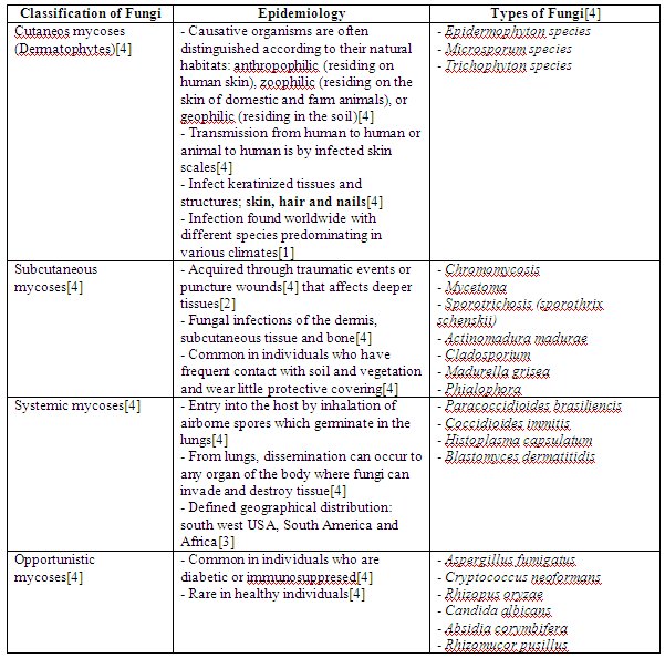
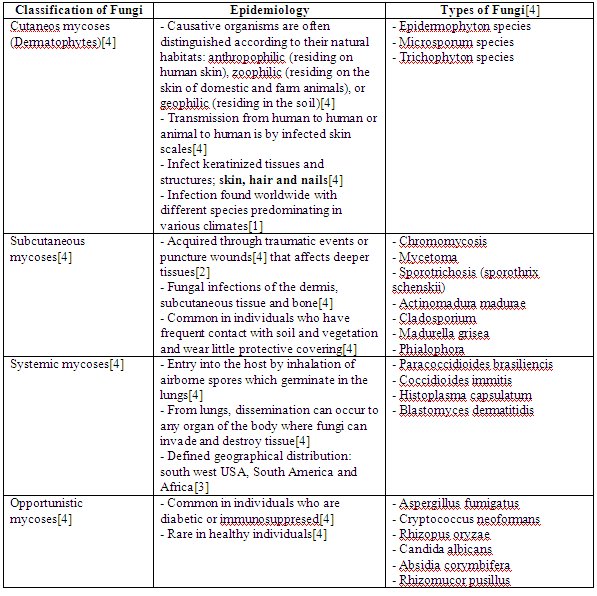
Medical Microbiology

Monday, January 29, 2007

dPBL2: Summarised table of commonly encountered fungal and parisite disease during jungal training

In our previous entry, we have listed down and explain about the possible fungal and parisital infection that may affect soldiers. For this entry, we have summarised the most common causes, which includes Tinea Pedis (athlete’s foot), Tinea Corporis (ringworm), Tinea Capitis(scalp ringworm),Tinea Cruris (jock’s itch), Tinea Unguim, Protozoa(Giardia Lamblia and Entamoeba Histolityca) and Helminths (Necatur Americanus[Hookworm] and Trichenia Trichuria [Whipworm]). This can be seen in the tables below:


References

[1] Richard A. Harvey, Pamela C. Champe and Bruce D. Fisher. Microbiology. 2nd edition. 2006. Lippincott Williams and wilkins
[2] Brooks, G. F., Butel, S. F., and Morse, S. A. (2004). Jawetz, Melnick & Adelberg’s Medical Microbiology (23rd ed.). Singapore: McGraw-Hill.
[3] http://wikipedia.org>
[4] http://www.apma.org/>
[5] http://www.umm.edu/>
[6] http://dermatology.about.com/>
[7] http://www.merck.com>
[8] Stephen Gillespie and Kathleen Bamford. Medical microbiology and infection at a glance. 2000. Blackwell science
[9] http://www.dpd.cdc.gov/>
[10] http://www.nlm.nih.gov/medlineplus/ency/article/001364.htm
[11] http://www.mayaparadise.com/>
[12] http://www.answers.com>

Sunday, January 21, 2007

Possible Fungal and Parasite infection on Soldiers

Fungus

There are many different classifications of fungus and parasites. The classification of fungi is summarized in the table below:


From the table above, the most possible cause of fungal infection for soldiers is cutaneous mycoses (dermatophytes). This is so as it is mainly caused by lack of hygiene practices, which may be common for soldiers.

In contrast, subcutaneous mycoses is more common for people who does plantation as it usually present in individuals who have frequent contact with soil and vegetation[4]. It is also unlikely that the soldiers can be infected by systemic mycoses as the defined geographical distribution excludes South East Asia countries. Opportunistic mycoses will also probably not infect soldiers as it is only common in individuals who are diabetic or immunosuppresed[4].

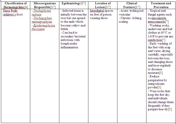
Now that dermatophytes is identified as the most possible cause of fungal infections for soldiers, we should then evaluate classification of this class of fungal infection. This can be summarized in the tables shown below:


From the table above, the main genres include Trichophyton, Microsporum and Epidermophyton.

Reasons how this fungus can infect soldiers:
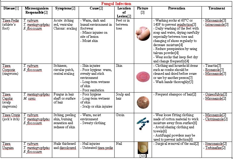
Tinea Pedis (athlete’s foot):
Soldiers will usually put on shoes for long hours each day. The fungus will most often attack the feet because shoes create a warm, dark, and humid environment, which encourages fungus growth [5]. If the feet are not washed adequately with soap and water, the fungus may continue to spread to the soles of the feet and to the toenails[5].

Tinea corporis (ringworm):
This fungus thrives in warm, moist areas. Poor hygiene, long-term wetness of the skin (such as from sweating) and minor skin and nail injuries usually raise the risk for a fungal infection. Tinea corporis is contagious when there is a direct contact with someone who is infected, or if in touch with contaminated items such as combs, clothing, shower floors and walls, or pool surfaces [11]. Soldiers usually experience poor sanitation and are usually in a warm, sweaty and sticky environment. Since it is contagious thus it will spread among the soldiers easily and by sharing contaminated items, it would increase the risk of the above fungal infection.

Tinea Cruris (“jock itch”):
Soldiers perspire often, creating a warm and moist environment for the fungus to grow. Causes of Tinea Cruris includes wearing sweaty or wet clothing[6]. Soldiers may also share clothes and towels. This is a possible cause of getting jock itch[6].

Tinea capitis (scalp ringworm):
An individual may have an increased risk for tinea infection if they have poor hygiene, prolonged wetness of the skin and minor skin or scalp injuries[11]. Prolonged wetness of the skin on the scalp may be due to the long period of time in wearing of the barret or helmet. Like Tinea corporis (ringworm), scalp ringworm are also contagious and a soldier may get infected if he come into direct contact with another soldier who has the condition, or by touching contaminated items such as combs, barret or clothing[11].

Tinea unguium (onychomycosis):
Onychomycosis is due to a fungal infection of the nail. Occasionally, fungal infection of the feet can lead to the nails becoming infected[20]. Tinea unguium often results from untreated tinea pedis(feet)[21]. When the soldiers encounter tinea pedis, it is usually left untreated for quite some time and this could result in the rise of tinea ungium.

Parasite
There are 3 main class of parasites that can cause infection to soldiers. This includes protozoa, helminths and scabies.

Protozoa
Protozoa can be classified based on the site of infection. This can be summarized in the table below:


* Amoebas: move by extending cytoplasmic projections
Flagellates: move by rotating whip-like flagella
Sporozoa: generally have non-motile adult forms

From the table above, it seems that Trypanosome species and Leishmania species can be ruled out as they are not commonly found in Asia. Plasmodium species can also been ruled out because based on our further research, Brunei has declared to be a malarial-free country, and thus it is least likely to have plasmodium species. Trichomonas vaginalis has also been ruled out as it is usually sexually transmitted. As for Cryptosporidium parvum, it has been rule out as well because it is more common when in direct contact with farm animals. As the cat (undercooked meat) is the definitive host for Toxoplasma gondii, it is ruled as well.

Thus the species that are possible for this situation would be Entamoeba histolytica, Giardia lamblia and Cyclospora cayetanensis.

Reasons how these protozoa can infect soldiers:

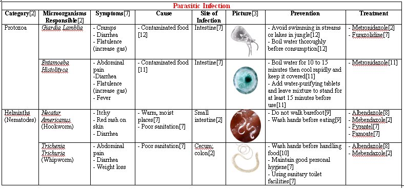
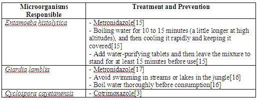
Entamoeba histolytica:
Entamoeba histolytica causes amoebic dysentery and is endemic in many tropical countries, but it is attributable more to unsanitary conditions than to heat[15].

Due to unsanitary conditions in the jungle, amoebic dysentery is most commonly spread by water or contaminated, uncooked food or from carriers. Flies may carry the cysts to spread the amoeba from the feces of infected persons to food[15].

Giardia lamblia (Giardia intestinalis):
Giardia lamblia
is found in soil, food, water, or surfaces that have been contaminated with the feces from infected humans or animals. It is common during travel to less developed countries in Southeast Asia[16].

With unsanitary conditions in the jungle, food might not be properly cooked and this could cause the transmission of Giardia lamblia. In addition, outbreaks among military personnel in the wilderness could also be due to an infection with various animal giardia harboured by animals such as rodents[17].

Cyclospora cayetanensis:
Cyclospora cayetanensis
is common in the tropics as warm and moist environment is essential before the oocyst sporulate into their infective form[13].

Transmission occurs in military personnel through the consumption of contaminated food and water[14].

The treatment and prevention of these microorganisms are summarised in the table below:


Helminths
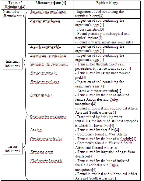
Helminths are worms, some of which are parasitic to humans[4]. The different types of helminths and microorganisms responsible are shown in the table below:


The epidemiology of the aboved microorganisms is summarised in the tables below. This is to exclude the microorganisms that is less possible to cause infection to soldiers.


From the table above, we have excluded most of the microorganisms and left with Necatur americanus (hookworm) and Trichenia trichuria (whipworm) as they are the most common microorganisms that could infect the soldiers.

Cestodes family also known as tapeworms are eliminated as they are due to undercooked meat (i.e: fish, pig, beef and etc.) and in this case we find that they seldom occur to the soldiers as they are often serve with food rations. Thus in this case, it is least likely to occur.

As for the Trematodes genre or also known as Flukes, they are also ruled out as based on their areas commonly found, none of them are commonly found in South East Asia. Thus, it is least likely to appear in Brunei jungle.

Nematodes genre or also known as the roundworms, only two species are specifically chosen. The other microorganisms are ruled out based on how they are transmitted or where they are commonly found.

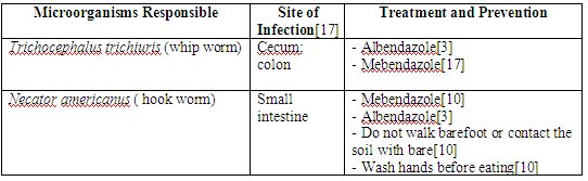
The site of infection, treatment and prevention of these helminths (hookworm and whipworm) are summarised in the table below:


Reasons how these helminths can infect soldiers:

Whip worm (Trichocephalus trichiuris):
Whipworm infection is very common in tropical areas where there is poor sanitation[12]. Warm climates provide the conditions for whipworm eggs to incubate in the soil[12].

Military personnel involved in jungle training most probably did not live in sanitary conditions and personnel could have acquired the parasite when they consume food that has been contaminated with eggs

Hookworm (Necator americanus):
Hookworm is most common in warm, moist places where sanitation is poor, such as the jungle. Two species of hookworm cause infection in people: Ancylostoma duodenale, which is present in India, China, Japan, and the Mediterranean area; and Necator americanus, which is present in the tropical areas of Africa, Asia, and the Americas[12]. Hence, the most likely species of hookworm infection commonly encountered by military personnel are of the Necator americanus species, as jungle training took place in Brunei.

Larvae emerge to live in the soil. When fully developed, the larvae can penetrate the skin. A person can become infected by walking barefoot or sitting in contaminated soil[12]. Infection may also occur when foods contaminated with hookworm cysts are eaten[8].

Hookworm eggs are passed in stool and infection results when a person comes in contact with the eggs from the contaminated soil, stool or water[9].

Military personnel are often protected against hookworms as they are fully clothed without any exposure of their skin. However, if their skin is exposed to soil during physical training, it would predispose them to hookworm infections as the larvae can penetrate through their skin.

Scabies
Scabies (Sarcoptes scabiei):
Scabies is a skin condition caused by a burrowing mite known as Sarcoptes scabiei[7]. It is a very contagious parasite which can spread easily through an infected individual through to other people. The infestation of this parasite is common, found worldwide, and affects people of all races and social classes[18]. It spreads more easily in crowded situations.

As army soldiers share the same tents, and are involved in many different physical activities, physical contact with one another would be a norm thus promoting the transmission of scabies throughout the entire population.

Treatment and Prevention:
- Avoid sharing common objects like clothing and bedding etc[7].
- Application of permethrin or oral dose of Invermectin for treatment[7].

References


[1] Tom Elliot, Mark Hastings and Ulrich Desselberger. Lecture notes on medical microbiology. 3rd edition. 1997. Blackwell science
[2] Medical microbiology lecture notes. Teamsek Applied Science School
[3] Stephen Gillespie and Kathleen Bamford. Medical microbiology and infection at a glance. 2000. Blackwell science
[4] Richard A. Harvey, Pamela C. Champe and Bruce D. Fisher. Microbiology. 2nd edition. 2006. Lippincott Williams and wilkins
[5] http://www.apma.org>
[6] http://dermatology.about.com>
[7] http://wikipedia.org>
[8] http://www.jhhs.org>
[9] http://www.personalmd.com>
[10] http://www.dpd.cdc.gov>
[11] http://www.nlm.nih.gov>
[12] http://www.merck.com>
[13] http://www.doh.state.fl.us>
[14] http://www.parasite-cleanse.com>
[15] http://www.mayaparadise.com/>
[16] http://www.answers.com>
[17] Brooks, G. F., Butel, S. F., and Morse, S. A. (2004). Jawetz, Melnick & Adelberg’s Medical Microbiology (23rd ed.). Singapore: McGraw-Hill.
[18] http://www.cdc.gov>
[19] http://www.umm.edu>
[20] http://www.ecureme.com>
[21] http://dermnetnz.org>

(The contents of this entry are contributed by all members. Tables are organised by Yasmin)

Thursday, December 14, 2006

3rd Entry - Summary of all Cases

All the necessary information is collated by all members and organised into tables. This can be seen below.


Abbreviations:
IMVic: indole-methyl red-Voges-Proskeur-Citrate
ONPG: O-nitrophenyl-β-D-galactopyranoside
OF: oxidation-fermentation
TSI: triple sugar iron
VP: Voges-Proskeur
MR: Methyl Red

References:
(1) http://findarticles.com/>
(2) http://medinfo.ufl.edu>
(3) http://tucom.bigbri.net/>
(4) http://en.wikipedia.org/>
(5) http://www.umm.edu/>
(6) http://textbookofbacteriology.net>
(7) Gabriel Virella. Microbiology and infectious diseases. Third edition
(8) http://w3.ouhsc.edu/>
(9)http://www.microbelibrary.org>
(10) http://members.tripod.com
(11) http://www.mc.maricopa.edu/>
(12) http://www.who.int/>
(13) http://www.rci.rutgers.edu/>
(14) G.Brooks, J.Butel and S.Morse. (2004). Jawetz, Melnick & Adelberg’s Medical Microbiology
(15) Henry D. Isenberg. Essential procedures for clinical microbiology

Friday, December 08, 2006

Culture media required for microorganism growth

As we will have to prepare the media culture for growing of the possible microorganisms, we have gathered information on the possible microorganisms and its corresponding appropriate culture media. This is summarised in the tables below.

For UTI cases (Patient 1,3 and 5) – Urine specimen [information gathered by Syafiqah & Samantha]


For enterocolitis case (patient 2 -Kwan Siew Lan ) [information gathered by Yasmin]


For food poisoning case (patient 4 - Ng Ming En) [8]
[information gathered by Farhana]


References:
[1] G.Brooks, J.Butel and S.Morse. (2004). Jawetz, Melnick & Adelberg’s Medical Microbiology
[2] http://medinfo.ufl.edu/year2/mmid/bms5300/bugs/klebpne.html
[3] http://en.wikipedia.org/wiki/Enterobacter
[4] http://medinfo.ufl.edu/year2/mmid/bms5300/bugs/candalb.html
[5] http://medinfo.ufl.edu/year2/mmid/bms5300/bugs/esccoli.html
[6] http://medic.med.uth.tmc.edu/path/bile.htm
[7] http://www.microbelibrary.org/ASMOnly/details.asp?id=2315&Lang
[8] http://www.findarticles.com/p/articles/mi_gGENH/is_/ai_2699003751

Thursday, December 07, 2006

Patient 1: Khong fay fay (2nd entry)

Reasons why cystitis was chosen for the diagnosis

1. Nausea and vomiting are signs and symptoms of pyelonephritis
a. Nausea and vomiting were not observed in this patient [1]

2. Causes of pyelonephritis include [1]
a. Urinary catherization[Kidney Infection (Pyelonephritis)
b. Surgery on urinary tract
c. Physical obstruction to the flow of urine due to a prostate enlargement or kidney stone or the backflow of urine from the bladder into the ureters increases the risk of pyelonephritis

However,
à Urinary catherization was not performed in this patient
à No history of surgery was indicated
à Patient is a female, hence physical obstruction to the flow of urine due to prostate enlargment is unlikely

3. Pyelonephritis is usually a spread of bacterial infection from the bladder [2]
à Risk of infection increases if there is a history of cystitis
à Hence even if pyelonephritis was present, cystitis is still the primary diagnosis

4. Pyelonephritis occurs less commonly in comparison to cystits. [3]

Complicated and Uncomplicated Cystitis
Cystitis is a common female problem. About 90% of UTIs in women are uncomplicated but recurrent. [4]

Cystitis is referred to as complicated if there are urologic abnormalities such as urinary retention, urinary stones and post surgery [5]

Bacteria found in cystitis
Uncomplicated cystitis
1. E. coli is the most commonly isolated microorganism in 80 - 90% of cystitis in young women followed by
2. Staphylococcus saprophyticus
3. Enterococci

Bacteria such as
1. Klebsiella
2. Proteus
3. Pseudomonads
constitute a lower percentage of infecting agents in uncomplicated cystits

Complicated cystitis
The spectrum of infecting bacteria in complicated cystitis is broader. Common infecting agents include
1. E. coli
2. Klebsiella
3. Proteus
4. Enterobacter
5. Enterococci
6. Pseudomonads
7. Staphylococci

Conclusion
Since uncomplicated cystitis is more common, the most probable bacteria that can be isolated from the bladder of this patient includes E. coli, Staphylococcus saprophyticus and Enterococci. Now that the common microorganisms have been identified, the next step is to find out ways to isolate the following bacteria and the biochemical tests that can be used for the identification of these bacteria.

References
1. http://www.merck.com/mmhe/sec11/ch149/ch149e.html
2. http://www.intelihealth.com/IH/ihtIH/WSIHW000/9339/10227.html
3. http://www.nlm.nih.gov/medlineplus/ency/article/000522.htm
4. http://www.faqs.org/health/Sick-V1/Cystitis.html
5. http://www.touchbriefings.com/pdf/1134/riedl.pdf
6. G.Brooks, J.Butel and S.Morse. (2004). Jawetz, Melnick & Adelberg’s Medical Microbiology

Patient 3: Maisy Wong (2nd entry)

Complaints (key words): Fever, chills, bladder distension. Patient is on indwelling catheter.

In this case, the urinary tract infection (UTI) can be caused by several reasons:

1) The patient is a 66 years old female. Hence the UTI may also be due to post menopause.
It is said that ‘the risk for UTI, both symptomatic and asymptomatic, is highest in women after menopause’ [2]. This may be due to bladder or uterine prolapse leading to incomplete bladder emptying [4]. Menopause may cause biologic changes. This in turn ‘put older women at particular risk for primary and recurring UTIs’ [2].

Post menopausal women will encounter estrogen loss. With this, the walls of urinary tract will thin out hence weakening the mucous membrane and reducing its ability to resist bacteria [2]. The bladder may also lose its elasticity, which eventually leads to failure to empty completely [2]. This points out to the cause of bladder distension for this patient.

Urethritis may occur in post menopausal women, whereby the tissues of the urethra and bladder become thinner and drier [5]. This brings about the infection.

Possible microorganisms: Klebsiella pneumoniae, Enterococci bacteria, E.coli and Proteus mirabilis.

2) Upper UTI brought by indwelling catheter (pyelonephritis)[1].
If a urinary catheter is left in place for a long time, bacteria will grow in it. A harmful infection may occur if the number of bacteria becomes large or if specific pathologic bacteria grow in the urinary tract [7].

Possible microorganisms: E. coli, Klebsiella and Proteus species, candida albicans.

3) Upper UTI caused by cystitis (lower UTI). Bacteria on catheter can infect the bladder, causing cystitis [3]. Although infection caused by catheter is associated with pyelonephritis, the bladder can also be infected as the catheter is inserted into the bladder.
When bacteria enter the bladder, they are usually eliminated through urination [6]. Infection will occur when bacteria multiply faster than they are removed by urination [6].

Older people have higher risk of developing cystitis due to incomplete emptying of the bladder associated with such conditions as benign prostatic hyperplasia (BPH), prostatitis and urethral strictures [6].

Possible microorganisms: E. coli, Staphylococcus saprophyticus, Klebsiella, Enterobacter and Proteus species.

I have researched on the morphology of the possible microorganisms. It has been collated together with Samantha's research. The table can be found in the entry below titled 'Morphology and Characteristics of Microorganisms in UTI'.

Now that I have listed the possible microorganisms for each posible cause of UTI, the next step is to find out the tests used in determining presence/absence of these microorganisms. This will be focused on in the next entry.

References:
[1] G.Brooks, J.Butel and S.Morse. (2004). Jawetz, Melnick & Adelberg’s Medical Microbiology
[2] http://adam.about.com/reports/000036_3.htm
[3] http://kidney.niddk.nih.gov/kudiseases/pubs/utiadult/
[4] http://www.emedicine.com/EMERG/topic626.htm
[5] http://scc.uchicago.edu/urethritis.htm
[6] http://en.wikipedia.org/wiki/Cystitis
[7] http://www.nlm.nih.gov/medlineplus/ency/article/000483.htm

Patient 4: Ng Ming En (2nd entry)

Patient: Ng Ming En
Diagnosis: Food Poisoning
Complaints: Severe vomiting, Diarrhea, Abdominal cramps

By looking at the symptoms associated with the different micro-organisms found in table in the previous entry and comparing them with the symptoms that the patient is experiencing, we can rule out Hepatitis A, Giardia Lamblia, Clostridium perfringens and Clostridium botulinum as the cause of the food poisoning.

The table below shows the remaining suspected micro-organisms and their microscopic morphologies.


References:
[1] G.Brooks, J.Butel and S.Morse. (2004). Jawetz, Melnick & Adelberg’s Medical Microbiology
[2] http://www.medscape.com/viewarticle/481644_print
[3] http://en.wikipedia.org/wiki/Rotavirus
[4] http://gsbs.utmb.edu/microbook/ch065.htm

Patient 5: Wong Wei Hong (2nd entry)

Wong Wei Hong
Male, 67 years old
Fever, chills, bladder distension; on indwelling catheter

The patient is an elderly man whose prostate is enlarged. This happens to all men as they grow older. As the gland grows, it presses on the urethra, the tube through which urine passes out of the body and causes urination and bladder problems, in this case, bladder distension.

As a treatment, catheter is inserted into the urethra to drain the urine as the patient has trouble emptying his bladder. The catheter, during it’s insertion through the urethra, might have displaced some of the normal flora into the bladder, causing urinary tract infection (pyelonephritis), the condition the patient is in now.

The suspected microorganisms are:
Escherichia coli (gram-negative bacilli)[1]
Klebsiella pneumoniae (gram-negative bacilli)[2]
Proteus mirabilis (gram-negative bacilli)[3]

A clean-catch urine should be collected for laboratory investigations. Urine should be cultured on a Columbia agar with 5% Sheep Blood (general purpose medium) and MacConkey agar (selective and differential medium used for the isolation of gram-negative organisms).[4]

[1]http://medinfo.ufl.edu/year2/mmid/bms5300/bugs/esccoli.html
[2]http://medinfo.ufl.edu/year2/mmid/bms5300/bugs/klebpne.html
[3]http://medinfo.ufl.edu/year2/mmid/bms5300/bugs/promirab.html
[4]www.nlm.nih.gov/medlineplus/ency/article/000522.htm

Morphology and Characteristics of Microorganisms in UTI

Morphology of microorganisms in Urinary Tract Infection (UTI)


References:
[1] G.Brooks, J.Butel and S.Morse. (2004). Jawetz, Melnick & Adelberg’s Medical Microbiology
[2] http://en.wikipedia.org/wiki/Klebsiella
[3] http://en.wikipedia.org/wiki/Enterococci
[4] http://en.wikipedia.org/wiki/Escherichia_coli
[5] http://tucom.bigbri.net/?p=108
[6] http://en.wikipedia.org/wiki/Enterobacter
[7]http://www.mycology.adelaide.edu.au/Fungal_Descriptions/Yeasts/Candida/Candida_albicans.html
[8] http://www.jyi.org/volumes/volume6/issue5/features/hu.html
[9] http://www.pdrhealth.com/drug_info/nmdrugprofiles/nutsupdrugs/pro_0034.shtml[10] http://medinfo.ufl.edu/year2/mmid/bms5300/bugs/esccoli.html
[10] http://medinfo.ufl.edu/year2/mmid/bms5300/bugs/esccoli.html

Friday, December 01, 2006

Patient 1: Khong Fay Fay

Urinary tract infection (UTI) refers to the infection of the urinary system which consists of the kidneys, ureter bladder and urethra. All of the components of urinary system can become infected but most infections involve the lower urinary tract that includes the bladder and the urethra. UTI occurs less commonly in men than in women. This is because the urethra in males is longer, making it difficult for bacteria to ascend the urinary tract. The urethra is the tube that carries urine from the bladder to the outside of the body.

There are 3 main types of UTI. These include:

Cystitis
§ Inflammation of the bladder which most commonly affects women, but can affect all age groups from either sex

Urethritis
§ Inflammation of the urethra which occurs equally in males and females; however, data may be skewed because urethritis is under recognized in women.

Pyelonephritis
§ Infection of the kidneys, which may occur after an infection in the bladder, has spreaded to the kidneys.


The incidence of these 3 types of UTIs may vary according to gender and across different age groups. The causes, signs and symptoms and incidence of these 3 types of UTI are summarized in the table below.


With regards to the case of Khong Fay Fay, the most possible diagnosis could be a UTI of the bladder, aka cysitis. Under the clinical diagnosis, there were complaints of fever, chills and dysuria. Dysuria refers to the painful or burning sensation that is felt during urination. In women, dysuria reflects inflammation of the bladder or the kidneys caused by a bacterial infection. It could also reflect inflammation of the vagina or vulva.

The patient is 26 years of age and could be sexually active. Hence, during sexual intercourse, bacteria could have been pushed up the urethra and easily into the bladder (since women have shorter urethra that allow bacteria to access the bladder more easily). This form of cystitis is also known as honeymoon cystitis.

If the patient is not sexually active, cystits could have resulted due to improper hygiene practices when women wipe from back to front. This would result in the introduction of normal flora such as E. coli from the anus to the urethra, leading to an opportunistic infection of the bladder and possibly the urethra. For her case, pyelonephritis is ruled out as symptoms of pyelnonephritis include a high fever, severe chills, nausea and vomitting which is not seen in this patient.

To further investigate the cause of UTI, midstream urine for microscopy and culture can be carried out. Test strips for blood, protein and nitrites may be used. The most common microorganims isolated is E. coli, but other bacteria such as proteus and Klebsiella pneumoniae which are normal flora of the bowels can be looked into. If the bacteria is transmitted during sexual intercourse, investigations for chlamydia and mycoplasma can be carried out.


References:
http://health.enotes.com/childrens-health-encyclopedia/cystitis
http://en.wikipedia.org/wiki/Cystitis
www.kidshealth.org/teen/sexual_health/stds/uti.html
http://www.students.stedwards.edu/kroduta
http://health.ivillage.com/bv/bvbladkidney/0,,7850,00.html
http://www.patient.co.uk/showdoc/40024549/

Patient 3: Maisy Wong

Case: Patient 3 -Maisy Wong

Diagnosis: Urinary Tract Infection

Complaints (Key words): Fever, Chills, Bladder distension. Patient is on indwelling catheter

In this entry, I will note down points on UTI in relation to the key words. I will then state down the possible microorganism(s) involved.

There are mainly 2 types of UTI – lower and upper. Hence we have to point out which of these 2 UTI is involved in this case. From here, we can then break down the possible microorganism(s) involved.

Bladder distension refers to the inability to urinate[1]. Catheterization is used to treat the bladder distension. Indwelling catheters avoid distension by emptying the bladder continuously into a bedside drainage collector[2].

One of the important keyword is that the patient is on indwelling catheter. As stated in the Jawetz, Melnick & Adelberg’s Medical Microbiology textbook (by G.Brooks, J.Butel and S.Morse), ‘Upper urinary tract infections routinely occur in patients with indwelling catheters even with optimal care and closed drainage systems’. Furthermore, fever and chills are the possible reactions after attachment of indwelling catheter. It was also mentioned in the textbook that ‘catherization carries a risk of introducing microorganism into the bladder, but it is sometimes unavoidable’.

The bladder wall is coated with various mannosylated proteins, which may interfere with the binding of bacteria to the uroepithelium[3]. The binding leads to establishment of pathogenicity for these organisms, its disruption results in reduced capacity for invasion of the tissues[3]. Furthermore, the unbound bacteria are more easily removed when voiding[3]. The use of physical trauma such as urine catheters may physically disturb this protective lining, thereby allowing bacteria to invade the exposed epithelium[3]. This paragraph explains how the introduction of indwelling catheter can lead to UTI.

From the above paragraphs, a possible diagnosis for the patient is upper urinary tract infection due to introduction of indwelling catheter. Hence the patient may have Pyelonephritis. The possible microorganism(s) involved are species of gram-negative rods. E. coli is however commonly found in UTI cases.

The next step in determining the possible microorganism(s) is to find out more about the gram-negative rods in relation to upper urinary tract infection. It is also important to find out more about the symptoms experienced by the patient and determine if the infection is due to before or after the catheter was introduced. Also, it is found that patient 5 also has the same complaints and diagnosis. However patient 3 is a female while patient 5 is a male. Hence, the difference have to be researched on.

Website References:
[1] http://www.wrongdiagnosis.com/medical/bladder_distension.htm
[2] http://www.healthatoz.com/healthatoz/Atoz/common/standard/transform.jsp?requestURI=/healthatoz/Atoz/ency/neurogenic_bladder.jsp
[3] http://en.wikipedia.org/wiki/Urinary_tract_infection

Patient 4: Ng Ming En

Case 4
Patient: Ng Ming En
Diagnosis: Food Poisoning


What is Food Poisoning?

A foodborne illness, also foodborne disease and commonly called food poisoning, is any illness resulting from the consumption of food contaminated by pathogenic bacteria, toxins, viruses, prions or parasites.

What are the common causative micro-organisms in Food Poisoning?


Since there is an extensive list of organisms that can cause Food Poisoning, the next thing we should do is to narrow down the list by looking at the symptoms present in the patient.

References:
http://www.ashtree.co.uk/features/bugs.htm
http://en.wikipedia.org/wiki/Food_poisoning

Patient 5: Wong Wei Hong

Using Samantha's table as a guide, I felt that Wong Wei Hong belongs to the third category (Pyelonephritis). The symptoms are as stated, fever and chills. On top of that, he also has urinary catherization.

Suspected organisms are all supposed to be normal, but displaced flora such as E.coli, Proteus sp and Klebsiella sp.